Anatomy Of A Microscope
A light microscope is a biology laboratory instrument or tool, that uses visible light to detect and magnify very small objects and enlarge them. They use lenses to focus light on the specimen, magnifying it thus producing an image. The specimen is normally placed close to the microscopic lens.
Parts of a Microscope Labeling Activity
With Labeled Diagram and Functions How does a Compound Microscope Work? Before exploring microscope parts and functions, you should probably understand that the compound light microscope is more complicated than just a microscope with more than one lens.

How to Use a Microscope (Properly) Step by Step New York Microscope
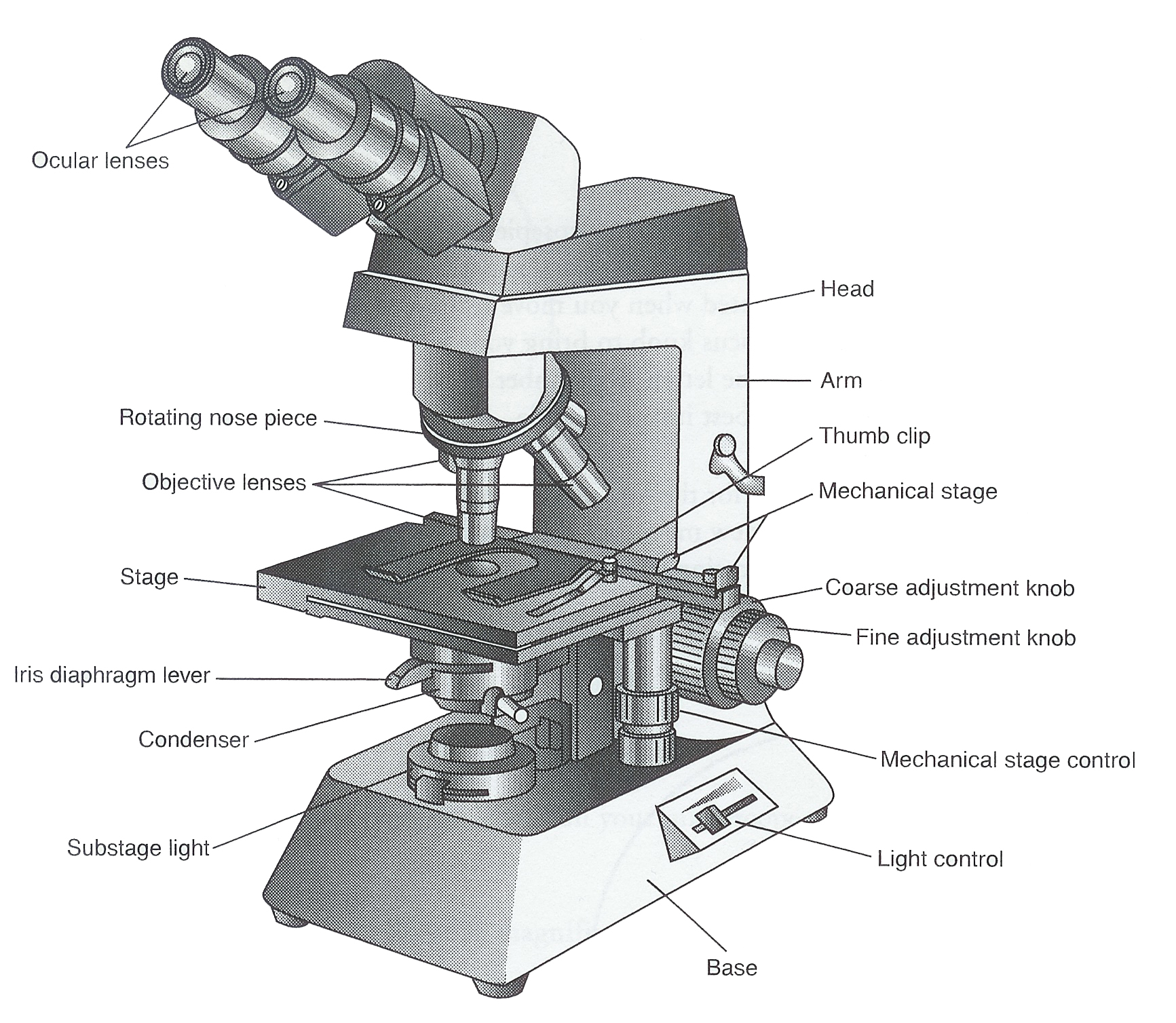
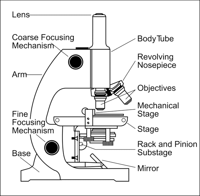
Major structural parts of a compound microscope. There are three major structural parts of a compound microscope. The head includes the upper part of the microscope, which houses the most critical optical components, and the eyepiece tube of the microscope.; The base acts as the foundation of microscopes and houses the illuminator.; The arm connects between the base and the head parts.
Parts Parts And Functions Of A Microscope
Parts Of a microscope. The main parts of a microscope that are easy to identify include: Head: The upper part of the microscope that houses the optical elements of the unit.; Base: The base is attached to a frame (arm) that is connected to the head of the device.The base of the microscope provides stability to the device and allows the user's hands to be free to manipulate other aspects of.

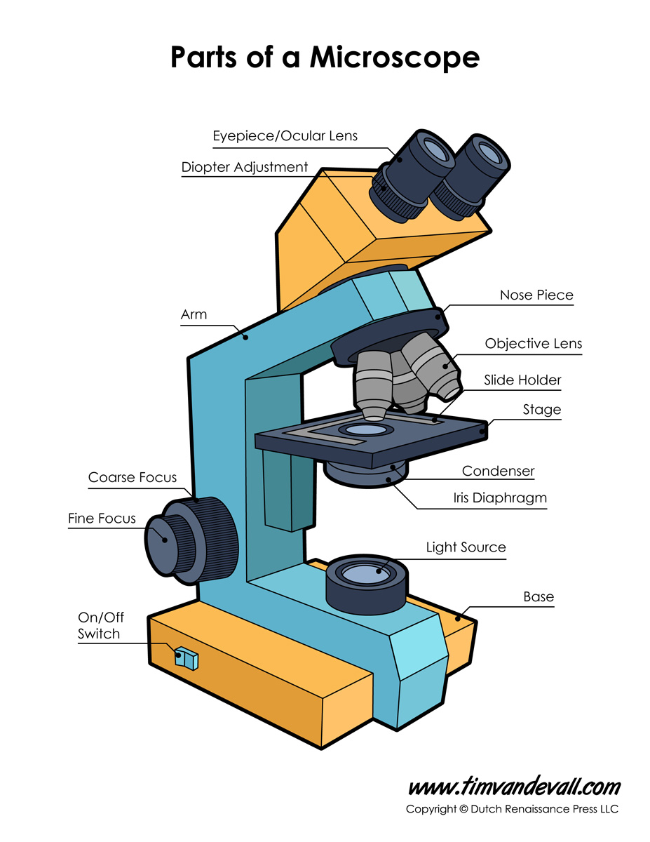
Labeled Microscope Diagram Tim's Printables
Magnification is a measure of how much larger a microscope (or set of lenses within a microscope) causes an object to appear. For instance, the light microscopes typically used in high schools and colleges magnify up to about 400 times actual size. So, something that was 1 mm wide in real life would be 400 mm wide in the microscope image.
Parts Of A Microscope With Functions And Labeled Diagram Images
Simple microscope is a magnification apparatus that uses a combination of double convex lens to form an enlarged, erect image of a specimen. The working principle of a simple microscope is that when a lens is held close to the eye, a virtual, magnified and erect image of a specimen is formed at the least possible distance from which a human eye.
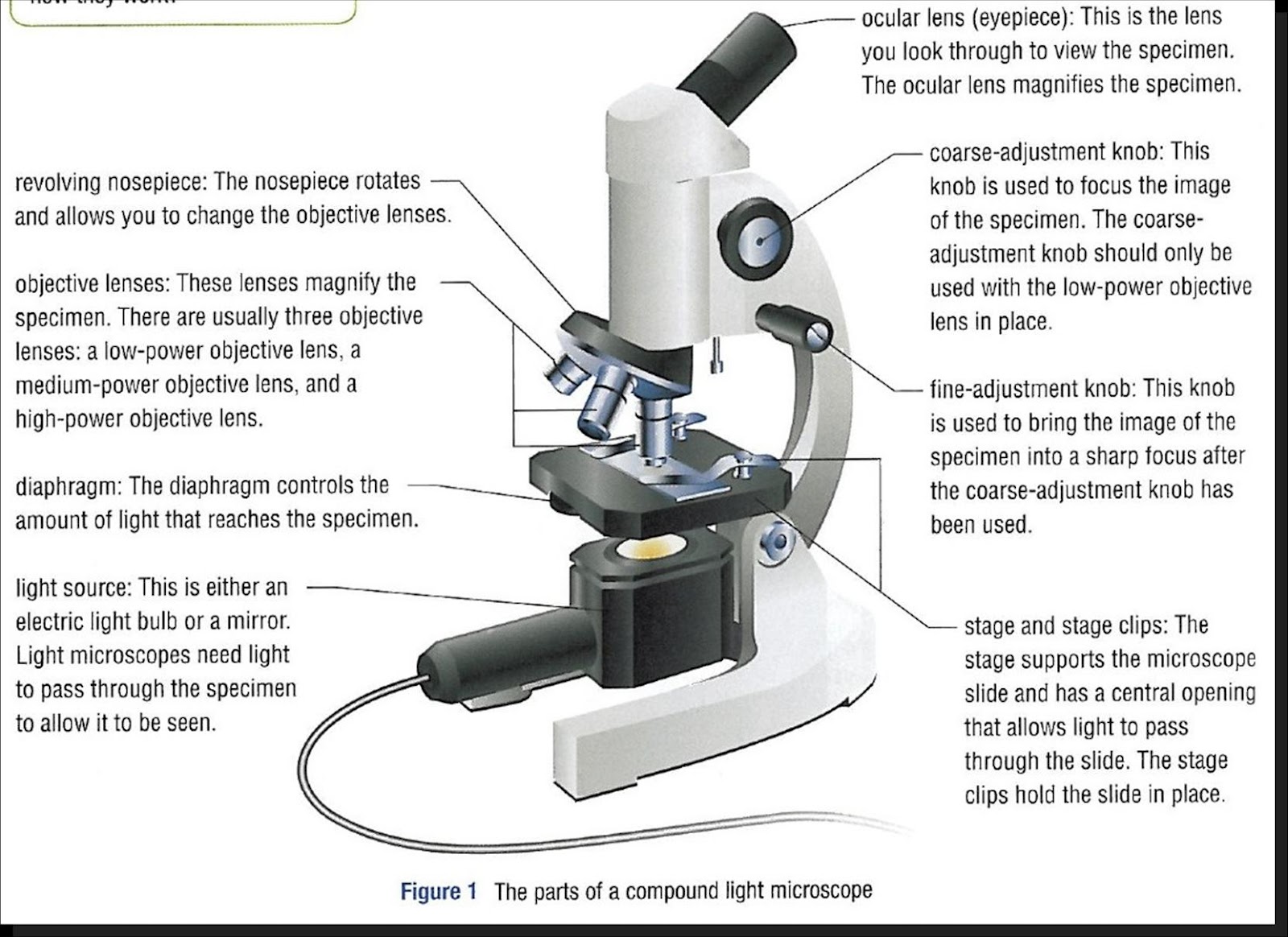
Monday September 25 Parts of a Compound Light Microscope
Explore the different parts of a microscope using a diagram, including the microscope lens, eyepiece, and stage. Updated: 10/13/2022 What is a Microscope? A microscope is a scientific.
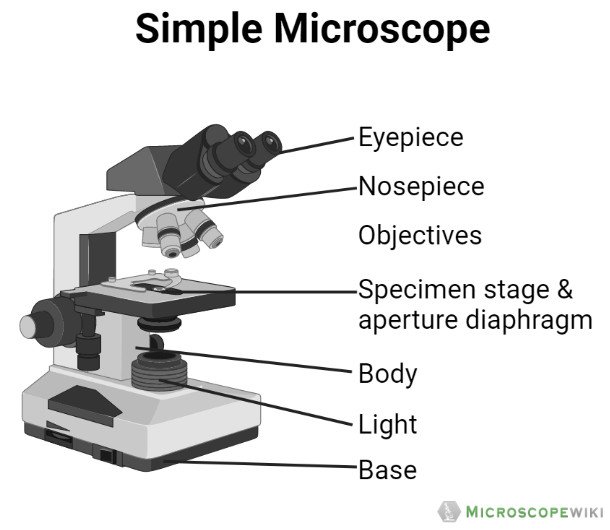
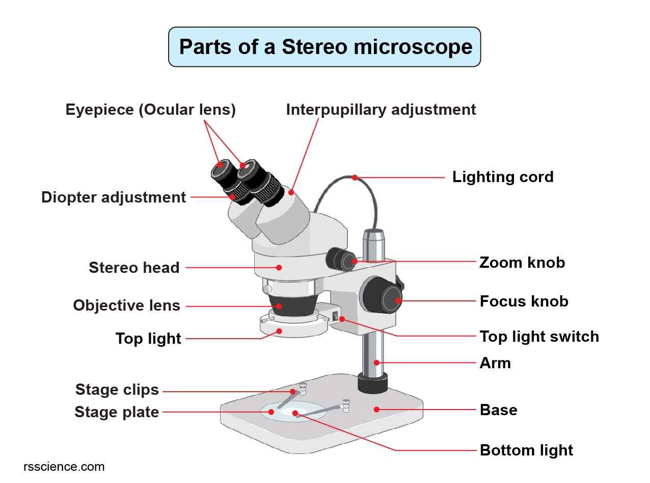
Parts of the Microscope (Labeled Diagrams) Simple and Compound Microscope
The hand magnifying glass can magnify about 3 to 20×. Single-lensed simple microscopes can magnify up to 300×—and are capable of revealing bacteria —while compound microscopes can magnify up to 2,000×. A simple microscope can resolve below 1 micrometre (μm; one millionth of a metre); a compound microscope can resolve down to about 0.2 μm.
1.5 Microscopy Biology LibreTexts
Structural parts of a microscope: There are three major structural parts of a microscope. The head comprises the top portion of the microscope, which contains the most important optical components, and the eyepiece tube.; The base serves as the microscope's support and holds the illuminator.; The arm is the component of the microscope that connects the eyepiece tube to the base of the.
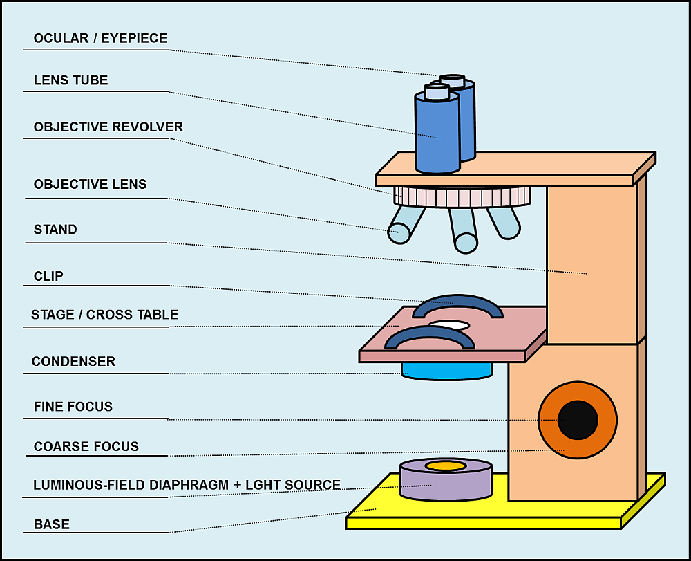
Parts and components of light microscopes Light Microscope
Body Tube Ocular Lens (eye-piece) Coarse and Fine Adjustment Knob Arm Base Microscope Worksheet The Light Microscope Light microscopes are used to examine cells at relatively low magnifications. Magnifications of about 2000X are the upper limit for light microscopes. The highest resolution of a light microscope is about 0.2 μm.

Parts of a microscope with functions and labeled diagram
2. Compound Microscope. Compound Microscope is a type of microscope that used visible light for illumination and multiple lenses system for magnification of specimen. Generally, it consists of two lenses; objective lens and ocular lens. It can magnify images up to 1000X.

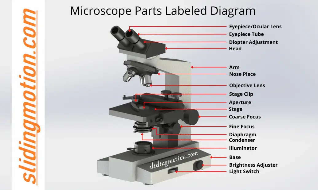
Microscope, Microscope Parts, Labeled Diagram, and Functions
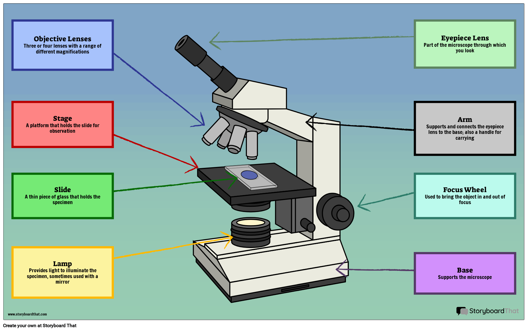
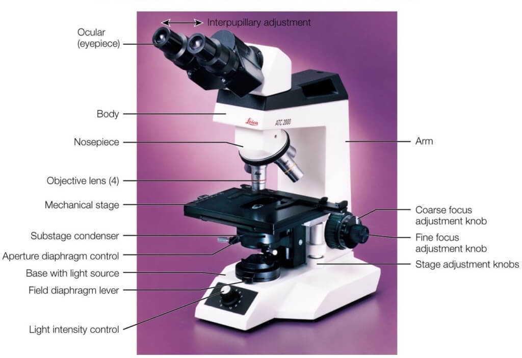
The individual parts of a compound microscope can vary heavily depending on the configuration & applications that the scope is being used for. Common compound microscope parts include: Compound Microscope Definitions for Labels Eyepiece (ocular lens) with or without Pointer: The part that is looked through at the top of the compound microscope.
Guide to understand microscope parts, names, functions & diagram
Parts of the Microscope (Labeled Diagrams) By Editorial Board December 14, 2022 The microscope is one of the must-have laboratory tools because of its ability to observe minute objects, usually living organisms that cannot be seen by the naked eyes. It is categorized into two: simple and compound microscopes.
Simple Microscope Definition, Principle, Parts, And Uses » Microscope Club
1. Head (Body) The head, also referred to as the body of the microscope, is a structural component that contains the optical parts of the microscope. The figure below shows the area of the microscope considered to be the body of the microscope.
Parts of a Microscope The Comprehensive Guide Microscope and
Q. Diagrammatically, identify the various parts of a microscope. Q. Describe the functions of each part of the microscope you have drawn above. Q. Differentiate between a condenser and an Abbe condenser. Q. What is the magnification power of the objective lenses? Q. How does the eyepiece compare to the objective lens? Q.
The Wonders Of Microscopes What You Need To Know Creyentes Diverses News
There are 1000 millimeters (mm) in one meter. 1 mm = 10 -3 meter. There are 1000 micrometers (microns, or µm) in one millimeter. 1 µm = 10 -6 meter. There are 1000 nanometers in one micrometer. 1 nm = 10 -9 meter. Figure 1: Resolving Power of Microscopes. The microscope is one of the microbiologist's greatest tools.